A pioneering research has produced the most comprehensive map of a mammalian brain ever documented.
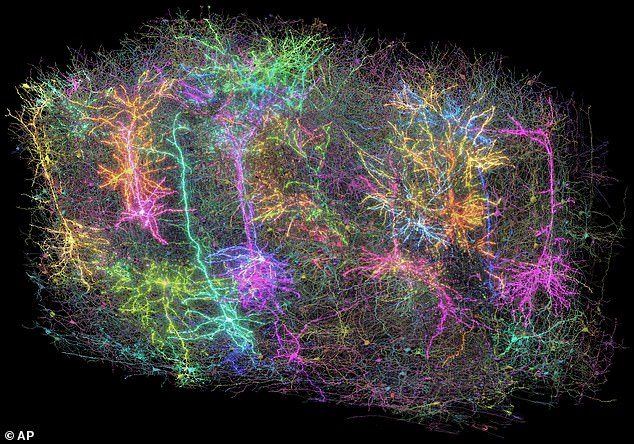
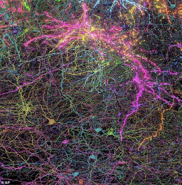
The 3D diagrams showcase over two miles of neuronal connections, nearly 100,000 neurons, and approximately 500 million synaptic junctions—all within a fragment of a mouse’s brain not larger than a grain of sand.
Dr. Clay Reid from the Allen Institute for Brain Science in Seattle stated, “Within this minuscule particle lies an intricate network of connections, governed by principles we have only just started to unravel.”
The specimen originates from an external section of the brain called the cortex, an area associated with vision.
Times
reports.
Dr. Forrest Collman from the same institute stated, “Through examining the functioning of the cortex in mouse brains, we can develop improved theories and hypotheses regarding the workings of our own brains.”
His group thinks that having the ability to chart and examine the brain’s inner connections at this scale could pave new avenues for understanding and addressing neurological disorders like
Alzheimer’s
disease, Parkinson’s and autism.
He referred to it as the ‘Google Maps for the brain,’ which goes beyond showing main highways to include each tiny street, individual houses, and even the rooms within those houses.
Similar to how individuals utilize Google Maps to determine the optimal path from location A to location B, or simply to verify if such a route is possible, this comprehensive neural map enables researchers to identify if two neurons are interconnected and precisely pinpoint where these connections take place.
What made this research particularly intriguing was that the mice needed their brain activity documented as they viewed YouTube videos, enabling researchers to observe the interactions between various clusters of neurons.
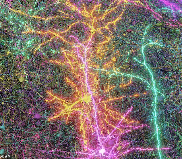
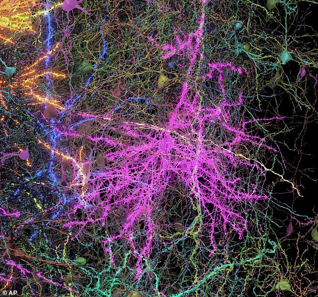
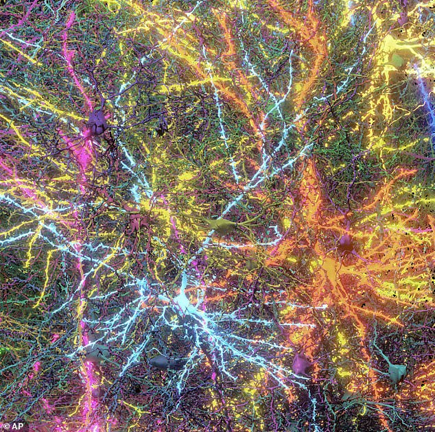
Following this, they cut the tissue into 25,000 slices, with each slice being merely 1/400th the thickness of a typical human hair, before examining these sections via powerful electron microscopes.
The images were combined to generate a 3D model with the help of artificial intelligence. The final result not only depicts the structure but also illustrates which brain cells interact and the manner in which they do so.
Nuno Macarico da Costa from the Allen Institute stated that one of the outcomes of their project reveals “just how extraordinarily beautiful the brain is.”
Simply observing these neurons allows you to grasp their complexity and size, instilling an overwhelming sense of wonder for the brain.
Read more